Medical Device History: Einthoven’s Triangle and the Electrocardiogram
Read time: 5 minutes
In our last blog, we summarized ISO 13485 and FDA regulations for the medical device manufacturing industry. This month, we will focus on the brief history behind Einthoven’s Triangle and the development of the electrocardiogram (ECG) – a game-changing medical device.
Along with other vital signs monitoring, electrocardiogram’s (ECG or EKG) aid medical professionals to detect and diagnose a patient’s potential life-threatening heart conditions. ECG is considered a vital sign and used primarily in emergency medical systems, hospitals and for mobile cardiac telemetry.
Who Created the Electrocardiogram(ECG)?

The first development of the electrocardiogram began with Dutch scientist, Willem Einthoven. Known as the “Father of ECG”, Willem Einthoven’s dedication to medical science led him to impactful discoveries in the medical field, such as, the ability to record heartbeats through Einthoven’s Triangle.
The String Galvanometer
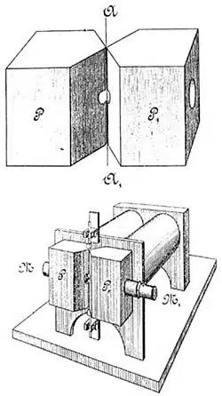
It was not until 1901 where Willem invented the string galvanometer. The string galvanometer is a medical device that allows for recording weak electrical pulses throughout the human body. The purpose of this medical device was for Willem to investigate the workings of the human heartbeat. The development of the string galvanometer and the discovery of the mechanism of electrocardiogram awarded Willem the Nobel Prize for Medicine or Physiology in 1924.
The Electrocardiogram
The string galvanometer plays an important role in the development of the electrocardiograph. In fact, the term, electrocardiograph was coined by Willem Einthoven, which in German translates to Elektro-kardiographie (EKG).
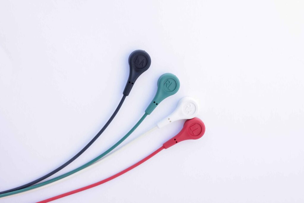
When applying an ECG on a patient, the snaps are patched and placed at certain parts of the chest, arms, and legs. These snaps which are referred to as electrodes and are connected to an ECG machine by lead wires. This creates electrical activity to diagnose heart activities and the ECG can record it. However, there is no electricity sent to the body. Instead, natural electrical impulses are recorded on different parts of the heart to maintain normal natural blood flow. Any changes in an ECG often signals potential heart-related conditions.
Einthoven’s Triangle Explained
Willem’s discoveries were such that cardinal limb leads form an equilateral triangle when deciding the electrical axis of the heart. The equilateral triangle formation connects to three limbs, the right arm, left arm, and left leg. This formation surrounds the heart into a circuit.
There are three lead systems that work together to use for the ECG which are:
- Standard Limb Leads
- Augmented Limb Leads
- Precordial Leads
Standard Limb Leads (bipolar) I, II, & III
These are used to show graph of potential difference between two limbs at a time where one limb carries a positive electrode, and the other limb carries a negative electrode. The three limb electrodes, I, II, and III form a triangle at the right arm (RA), left arm (LA), and left leg (LL).
In mathematical terms, Einthoven’s Law Explains that Lead II’s complex is equal to the sum of the corresponding complexes in Leads I and III. (II = I + II)

Augmented Limb Leads (unipolar) aVR, aVL, & aVF
These are used to show a graph of recorded electrical forces one limb at a time. The leads, aVR, aVL, and aVF can read more information about the heart because it is placed in three more directions on the frontal plane.

Precordial Leads: V1-V6
Lastly, there are precordial leads. Also known as “Chest Leads”, they are placed on the sternum in a posterior (toward the back) direction. These leads are lined in a horizontal direction, where more of the hearts signal can be viewed alongside the earlier limb lead.

The String Galvanometer and Electrocardiograms Continued
The intentionality of the string galvanometer continued to develop as more scientists began to further study the function and diseases of the heart muscles. The laboratory at Leiden then became a scientific landmark for all scientists around the world.
Here’s a picture to compare the earlier designs versus the most current design of ECG machines:


Although EKGs are used across the globe, there are a few defining factors that visually differentiate separate American and European standards. These standards come from American Heart Association (AHA) and International Electro-technical Commission (IEC). These two standards have been developed and produced in the early 90’s at two different time and although many revisions have been made, the standards remain separate.
Below is a simple chart to show AHA and IEC differences:

Conclusion:
Willem’s scientific impact paved the way for the medical industry to develop what ECGs are now today. The use of Einthoven’s Triangle is still an important reference for developers as ECG’s continue to evolve. Without Willem’s discoveries, where would the medical industry be? Would the development for current ECG’s have progressed as they did?
The team at ClearPath Medical has decades of experience designing, and manufacturing high quality ECG patient leads and cable assemblies, compliant to ANSI AAMI EC53 and IEC 60601 requirements for medical original equipment manufacturers. From concept to full-scale production, including design verification testing, ClearPath Medical excels in bringing robust, effective solutions for critical, life-saving medical cable assemblies.
Thanks for reading! Here are some more ClearPath Medical blogs that might interest you.
Love what you see? Sign up for our newsletter to get the latest updates on our company, worldwide and more.
Sources:
-
-
John Hopkins Medicine. “Electrocardiogram” Health, 2022, Accessed October 27, 2022. https://www.hopkinsmedicine.org/health/treatment-tests-and-therapies/electrocardiogram#:~:text=Electrodes%20(small%2C%20plastic%20patches%20that,is%20sent%20into%20the%20body
-
Medical Design & Outsourcing. “MedTech Memoirs: The Electrocardiograph (EKG)” September 16, 2015, Accessed October 27, 2022. https://www.medicaldesignandoutsourcing.com/medtech-memoirs-the-electrocardiograph-ekg/
-
National Library of Medicine. “On the Einthoven Triangle: A Critical Analysis of the Single Rotating Dipole Hypothesis” PubMed Central, July 20, 2018, Accessed October 27, 2022. https://www.ncbi.nlm.nih.gov/pmc/articles/PMC6068749/
-
The Physiologist. “The ECG Leads, Polarity and Einthoven’s Triangle” The Student Physiologist, 2017, Accessed October 27, 2022.
https://thephysiologist.org/study-materials/the-ecg-leads-polarity-and-einthovens-triangle/
-